By David A. Goldman MD –
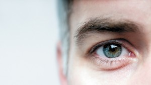
 Although people may have heard the word ‘cornea’ in reference to the eye, many are unaware of what is really refers to. While diseases such as glaucoma and macular degeneration affect the back of the eye, the cornea is in the front of the eye.
In fact, the cornea is the most anterior structure of the eye. It is the clear part responsible for much of the focusing of light waves which enter the eye. When patients place contact lenses on their eye, they are placing them on their corneas. When patients develop an abrasion (scratch) or infectious ulcer of the eye, it is typically in the cornea.
The cornea consists of several layers, but it is simplest to break it down into three: epithelium, stroma, and endothelium. The epithelium is the superficial outer layer of cells. When a scratch occurs on the cornea, the epithelium acts quickly to fill in the scratch. In cases of severe dry eye, tiny dried out “holes” can also appear in the epithelium.
The stroma is the central portion of the cornea, and compromises the bulk of it. It provides the majority of structural support to the cornea. When patients undergo LASIK surgery, it is the stroma that is affected. In LASIK, a laser creates a flap within the stroma. That flap is then lifted and a laser then reshapes the cornea under the flap. For patients who are myopic (nearsighted), the laser flattens the center of the cornea. For patients who are hyperopic (farsighted), the laser removes tissue in a circular pattern to steepen the cornea.
The endothelium is the most posterior layer of the cornea. Consisting of a layer of cells, these endothelial cells work as microscopic pumps to pump fluid out of the cornea to keep it clear. As we age the number of endothelial cells decline. Typically this is asymptomatic. In conditions such as Fuchs Dystrophy, however, patients are born with a lower number of endothelial cells and at some point may require surgery. Fortunately, the surgical options have improved greatly – less than ten years ago a new procedure was developed called DSAEK. In the past, corneal swelling which did not respond to topical eye drops required a full thickness corneal transplant. With the DSAEK procedure, solely the posterior layer of cells is transplanted. With this newer technique, vision can be restored in weeks instead of months.
This is not to say that a full thickness corneal transplant does not have its place in eye care. Conditions such as keratoconus, where the cornea becomes irregularly cone shaped, and scars of the cornea can significantly limit visual acuity. In these cases, replacing all layers of the cornea can work well to restore vision. In many cases, laser vision correction can be performed over a corneal transplant so that the patient can see well without glasses.
While problems of the cornea, whether inherited of environmental, can significantly disturb vision, there are multiple procedures available to improve vision. As opposed to posterior eye pathology such as glaucoma and macular degeneration, the overwhelming majority of vision problems related to the cornea can be fixed.
Prior to founding his own private practice, Dr. David A. Goldman served as Assistant Professor of Clinical Ophthalmology at the Bascom Palmer Eye Institute in Palm Beach Gardens. Within the first of his five years of employment there, Dr. Goldman quickly became the highest volume surgeon. He has been recognized as one of the top 250 US surgeons by Premier Surgeon, as well as being awarded a Best Doctor and Top Ophthalmologist.
Dr. Goldman received his Bachelor of Arts cum laude and with distinction in all subjects from Cornell University and Doctor of Medicine with distinction in research from the Tufts School of Medicine. This was followed by a medical internship at Mt. Sinai – Cabrini Medical Center in New York City. He then completed his residency and cornea fellowship at the Bascom Palmer Eye Institute in Miami, Florida. Throughout his training, he received multiple awards including 2nd place in the American College of Eye Surgeons Bloomberg memorial national cataract competition, nomination for the Ophthalmology Times writer’s award program, 2006 Paul Kayser International Scholar, and the American Society of Cataract and Refractive Surgery (ASCRS) research award in 2005, 2006, and 2007. Dr. Goldman currently serves as councilor from ASCRS to the American Academy of Ophthalmology. In addition to serving as an examiner for board certification, Dr. Goldman also serves on committees to revise maintenance of certification exams for current ophthalmologists.
Dr. Goldman’s clinical practice encompasses medical, refractive, and non-refractive surgical diseases of the cornea, anterior segment, and lens. This includes, but is not limited to, corneal transplantation, microincisional cataract surgery, and LASIK. His research interests include advances in cataract and refractive technology, dry eye management, and internet applications of ophthalmology.
Dr. Goldman speaks English and Spanish.
561-630-7120 | www.goldmaneye.com
Check Also
WHY DO I FEEL SO TIRED ALL OF THE TIME, AND NOTHING WAKES ME UP?
By Renee Chillcott, LMHC When it comes to a feeling we can’t tolerate and want …
 South Florida Health and Wellness Magazine Health and Wellness Articles


