 South Florida Health and Wellness Magazine Health and Wellness Articles

Related Articles

By Lauren R. Rosecan, M.D., Ph.D., F.A.C.S. –
 Histoplasmosis is a disease caused when airborne spores of the fungus Histoplasma capsulatum are inhaled into the lungs. This microscopic fungus, sometimes called histo for short, is released into the air when soil is disturbed by plowing fields, sweeping chicken coops, or digging holes.
Histoplasmosis initially is a lung infection. However, it is believed that the infection, even if mild, can later migrate to the eye through the blood stream and cause a serious eye disease called ocular histoplasmosis syndrome (OHS). OHS is a leading cause of vision loss in Americans ages 20 to 40.
Histoplasmosis Symptoms
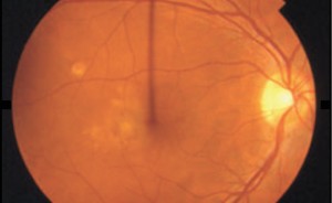
Ocular histoplasmosis syndrome (OHS) often has no symptoms in its early stages. You may have been affected by OHS without knowing it. The evidence that the inflammation ever occurred are tiny scars called “histo spots,” which remain at the infection sites. Histo spots do not generally affect vision, but for reasons that are still not well understood, they can result in complications years — sometimes even decades — after the original eye infection. Histo spots have been associated with the growth of the abnormal blood vessels underneath the retina.
In later stages, histoplasmosis symptoms may appear if the abnormal blood vessels cause changes in vision. The symptoms for OHS are the same as choroidal neovascular membrane symptoms .
These include:
• Blank spots in your vision, especially your central vision;
• Distorted vision, so that straight lines appear bent, crooked or irregular;
• Size of objects may appear different for each eye;
• Colors lose their brightness; colors do not look the same for each eye;
• Central light flashes or flickering.
How Is Histoplasmosis Diagnosed?
Your Eye M.D. will be looking for two things in particular:
• The presence of histo spots, which indicate previous exposure to Histoplasma capsulatum fungus spores;
• Swelling of the retina, which signals the growth of new, abnormal blood vessels.
The examination to diagnosis histoplasmosis is similar to that used for a wet macular degeneration diagnosis. Your doctor may have you use an Amsler grid to check for histoplasmosis symptoms such as wavy, blurry or dark areas in your vision.
As part of the examination, your Eye M.D. will dilate (widen) your pupils using dilating eyedrops and examine your eyes with an ophthalmoscope, a device that allows him or her to see the retina and other areas at the back of the eye. If fluid or abnormal blood vessels (choroidal neovascular membranes) are detected, your ophthalmologist will take special photographs of your eye with optical coherence tomography (OCT) and fluorescein angiography.
OCT scanning uses light waves to create detailed images of the underlying structure of the retina. OCT images show the thickness of the retina, and can help your Eye M.D. detect swelling and abnormal blood vessels.
During fluorescein angiography, a fluorescein dye is injected into a vein in your arm. The dye travels throughout the body, including your eyes. Photographs are taken of your eye as the dye passes through the retinal blood vessels. Abnormal areas will be highlighted by the dye.
How Is Histoplasmosis Treated?
Anti-VEGF treatment
One method for treating histoplasmosis targets a specific chemical in your body that causes abnormal blood vessels to grow under the retina. That chemical is called vascular endothelial growth factor, or VEGF. Several new drug treatments (called anti-VEGF drugs) have been developed that can block the trouble-causing VEGF. Blocking VEGF reduces the growth of blood vessels, slows their leakage, helps to slow vision loss, and in some cases improves vision.
Your ophthalmologist (Eye M.D.) administers the anti-VEGF drug directly to your eye in an outpatient procedure. Before the procedure, your ophthalmologist will clean your eye to prevent infection and will use an anesthetic to numb your eye with a very fine needle. You may receive multiple anti-VEGF injections over the course of many months. Repeat anti-VEGF treatments are often needed for continued benefit.
Laser treatment
Laser treatment for histoplasmosis is usually done as an outpatient procedure in the doctor’s office or at the hospital.
The laser beam in this procedure is a high-energy, focused beam of light that produces a small burn when it hits the area of the retina to be treated. This destroys the abnormal blood vessels, preventing further leakage, bleeding and growth.
Following laser treatment, vision may be more blurred than before treatment, but often it will stabilize within a few weeks. A scar forms where the treatment occurred, creating a permanent blind spot that might be noticeable in your field of vision.
Laser treatment does not cure histoplasmosis. However, it reduces the chance of abnormal blood vessels returning. If these blood vessels do return, additional laser surgery may be needed.
Steroid injection
Because histoplasmosis can cause inflammation in the eye, sometimes steroid injections are given in the eye to reduce the swelling.
Histoplasmosis remains a threat to your vision for your lifetime. Therefore, it is important to have regular checkups with your ophthalmologist to detect any problems as early as possible.
Lauren R. Rosecan, M.D.
West Palm Beach
901 North Flagler Drive, 33401
(561) 832-4411 Office
(561) 832-1591 Fax
Stuart
618 East Ocean Blvd., #3, 34994
(772) 287-7026 Office
(772) 220-4186 Fax
Palm Beach Gardens
11382 Prosperity Farms Rd., #128, 33410
(561) 627-7311 Office
(561) 627-6791 Fax
Boca Raton
1050 NW 15th Street, #114, 33486
(561) 368-7723 Office
(561) 368-0093 Fax
